TheJakartaPost
Please Update your browser
Your browser is out of date, and may not be compatible with our website. A list of the most popular web browsers can be found below.
Just click on the icons to get to the download page.

Popular Reads
Top Results
No results found. Please check your search term and try again
Can't find what you're looking for?
View all search resultsPopular Reads
Top Results
No results found. Please check your search term and try again
Can't find what you're looking for?
View all search resultsNew technique opens window into how brain cells communicate
Change text size
Gift Premium Articles
to Anyone
Share the best of The Jakarta Post with friends, family, or colleagues. As a subscriber, you can gift 3 to 5 articles each month that anyone can read—no subscription needed!
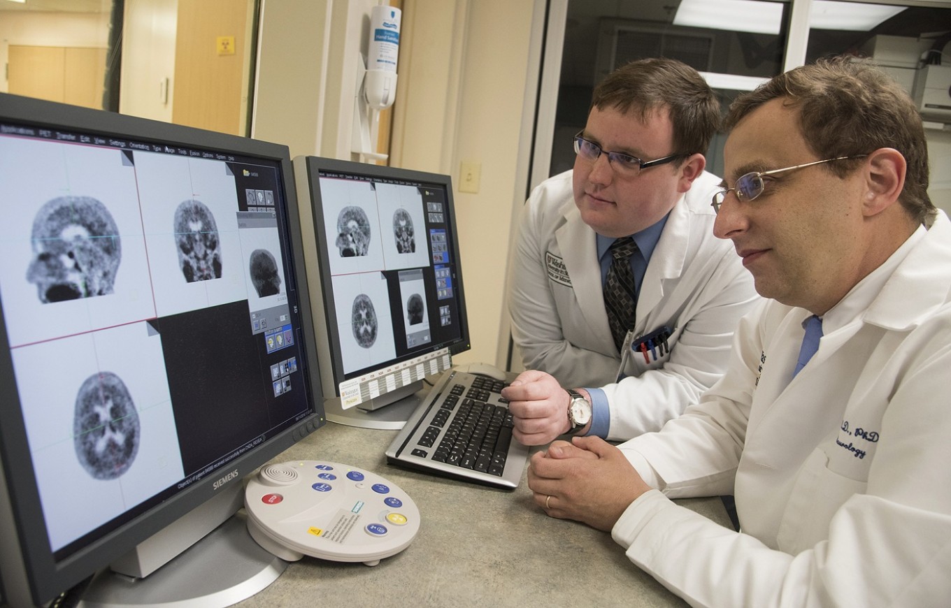 This photo provided by Washington University shows associate professor of neurology at Washington University School of Medicine Beau Ances MD, PhD, right, and Matthew Brier an MD/PhD student at the university, examining PET (positron emission tomography) scans of Alzheimer’s disease patients, in St. Louis. (Washington University via AP/Robert Boston)
T
he brain's nerve cells communicate by firing messages to each other through junctions called synapses, and problems with those connections are linked to disorders like Alzheimer's and epilepsy. Now Yale University researchers have developed a way to picture synapses in living brains.
The technique reported Wednesday, using PET scans, is highly experimental but it raises the possibility of one day monitoring synapse function in some common diseases.
A healthy human brain harbors trillions of synapses, a number that changes over a lifetime.

Early in life, the brain "prunes" the many synapses between neurons so the right number is in each region, a process that can go wrong in disorders such as autism or schizophrenia. Changes in the density of synapses may signal where epilepsy seizures originate. Later in life, synapse loss is associated with Alzheimer's disease.
But measuring synapses has required autopsies, or occasional attempts during brain surgery.
To find a non-invasive approach, the Yale-led team developed a radioactive compound, called a tracer, that is injected into the body and binds with a particular protein that is found in the brain's synapses. The idea: During a PET scan, those synapses appear lit up against dark, synapse-free areas of the brain.
Animal testing confirmed the tracer was targeting synapses.
(Read also: Study: Brain scans reveal hidden consciousness in patients)
 In this photo provided by Science Translational Medicine, PET scans taken at the Yale PET Center show the density of connections between nerve cells, called synapses, in a healthy living brain. Yale researchers developed a way to picture synapses in a living brain, something that until now has been studied mostly during autopsies. (Science Translational Medicine via AP/-)
The research team then mapped the density of synapses in the brains of 10 healthy volunteers and three patients with a form of epilepsy. Compared to the healthy brains, the technique revealed lost synapses in the epilepsy-affected regions of those patients' brains, the researchers reported Wednesday in the journal Science Translational Medicine.
"This work represents a breakthrough in the ability to study an important process in the brain that is not only part of normal brain development but that also may be involved in several neuropsychiatric diseases," said Dr. Peter Herscovitch, who directs PET scanning at the National Institutes of Health's Clinical Center and wasn't involved in the research.
Much more work is needed to make the tracer last longer in the brain, a key if it's ever to be of use to doctors, cautioned Yale radiology professor Richard Carson, the study's senior author. But even though it starts disappearing quickly, he said it's a good tool to research brain function.
Stay tuned: Carson's team has begun using the technique to study Alzheimer's, to determine if changes in synaptic density over time can help predict that disease's development.