TheJakartaPost
Please Update your browser
Your browser is out of date, and may not be compatible with our website. A list of the most popular web browsers can be found below.
Just click on the icons to get to the download page.

Popular Reads
Top Results
No results found. Please check your search term and try again
Can't find what you're looking for?
View all search resultsPopular Reads
Top Results
No results found. Please check your search term and try again
Can't find what you're looking for?
View all search resultsFujifilm integrates AI technology with health services solutions
Change text size
Gift Premium Articles
to Anyone
Share the best of The Jakarta Post with friends, family, or colleagues. As a subscriber, you can gift 3 to 5 articles each month that anyone can read—no subscription needed!
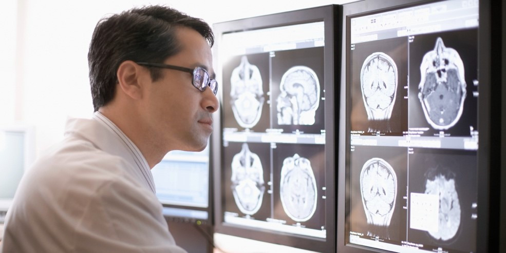 A health worker reads a brain scan in this handout photo. (Courtesy of Fujifilm Indonesia)
Fujifilm, the imaging, information and document solutions giant, has been diversifying its business for a long time.
One of the business lines that continues to develop and record profits is its health services technology business, which combines Fujifilm’s sophisticated imaging and filmmaking technologies with the diagnostic equipment needed by the healthcare industry.
Fujifilm Indonesia President Director Masato Yamamoto said quality medical technology was vital to help health workers provide examination services and diagnose diseases to make the right decisions for their patients.
"We hope that everyone can receive high-quality medical care," said Yamamoto.
While it is primarily known for its photography equipment and services business, Fujifilm’s engagement in the healthcare industry spans nearly 90 years. The diversified conglomerate started out as a medical imaging specialist in 1936, selling X-ray film.
With such a legacy, the company’s leadership is well aware of the technological advancements in film manufacturing and imaging processes, as well as the growing market demand for these technologies and related solutions. In the healthcare industry, advanced technologies are invaluable in medical diagnostic and imaging equipment.
Approaching these conditions as an opportunity for further growth, Fujifilm has expanded its diagnostics business by applying artificial intelligence (AI) to its array of medical equipment, such as X-ray and ultrasound imaging, endoscopes, in vitro diagnostic systems, computerized tomography (CT) scans and magnetic resonance imaging (MRI), as well as picture archiving and communication systems (PACS).
According to Yamamoto, Fujifilm realizes that clinical data will be increasingly necessary to maintain health and sustain life in the future. For this reason, the company is expanding its use of AI in developing products that meet the growing needs of the healthcare industry, especially in the era following the global COVID-19 pandemic.
Fujifilm has been collaborating with many parties on researching the potential development and use of AI in medicine.
In April 2019, for example, Fujifilm announced that it had successfully codeveloped with Kyoto University an AI-based technology to automatically identify and measure interstitial pneumonia lesions with higher precision. The technology was developed from a Fujifilm-Kyoto University study that used AI to help diagnose patients with COVID-19 pneumonia and assess their treatment.
Fujifilm's other initiatives include developing an AI platform for use in both emergency rooms and operating rooms, including for X-ray imaging during general checkups.
One of the company’s visions it to provide technological support to radiographers, radiologists and surgeons during an operation. As part of its efforts to fulfill this vision, Fujifilm has developed medical systems employing medical information technology (IT) solutions that can be combined with its wide array of diagnostic and imaging products.
Mammography technology
Medical systems and equipment incorporating advanced technology comprise the main portfolio in Fujifilm's business transformation.
In July 2019, Fujifilm launched in Japan its integrated medical software system, an AI-powered platform that employs machine learning to use the knowledge of experienced doctors in medical image analysis and diagnosis.
Drawing on its industry expertise of more than 20 years and technological sophistication, Fujifilm has also developed an AI-integrated medical imaging and information management system called Synapse PACS, which is capable of managing and storing imaging data at hospitals around the world. Launched in 1999 as the first web-based PACS in the world, Synapse PACS is now used by 5,500 health facilities around the globe.
Further, Fujifilm is using its imaging and processing technologies as the basis for future development of its AI technology.
According to Yamamoto, Fujifilm's IT experts analyze data from the company's high-quality medical imaging solutions as in-depth learning material to produce other AI and imaging innovations.
He also emphasized that one of Fujifilm's was how patients could get immediate and accurate medical treatment through diagnoses based on X-ray images, such as from chest X-rays or mammography.
One of the sophisticated medical imaging equipment from Fujifilm is a high-resolution mammography, developed as part of the company’s “Never Stop” campaign, on “innovating solutions for a better world”.
Breast cancer is the second leading cause of global deaths from cancer among women. Therefore, early detection of breast cancer from mammography will have a significant impact on the life expectancy and quality of life of a patient.
Detecting breast cancer usually involves a doctor or oncologist advising a patient to get an X-ray examination so they can take the necessary actions.
This is what happened to Anjani, a social media content specialist who was diagnosed with breast cancer. In the beginning of 2021, Anjani felt a strange lump in her right breast. She did not worry about it at the time, as she had a mammography every year and the results were always negative. However, 2020 was the one year she didn’t have a mammography because of the COVID-19 pandemic.
As time passed, however, she felt that something was wrong. Finally, she saw an oncologist, who referred her to get a mammography of the breast glands and surrounding tissue to check whether the unusual growth was benign or malignant.
After her mammogram was analyzed, the oncologist determined that the breast lump was malignant and that she needed oncological surgery, specifically a mastectomy to remove her right breast.
Anjani's experience illustrates how a mammography can accurately locate and identify malignant cancer cells in a patient’s body. Analyzing the resulting mammogram can help doctors diagnose breast cancer and give a “second life” to their patient by determining the appropriate medical action and treatment, before it is too late.
This is where Fujifilm's mammography technology comes in, by contributing to the early detection of breast cancer. Fujifilm's mammography can assist medical professionals in the early detection of anomalous tissue growth in a patient's body. They can then provide a correct and accurate diagnosis for recommending the appropriate procedure and treatment.
Reducing the burden of radiation on a patient during a mammography is one of the reasons Fujifilm continues to improve its technology, by leveraging its knowledge of X-ray imaging and image processing technology it has developed for almost 90 years.
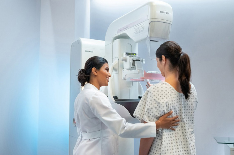
"Fujifilm continues to develop its mammography technology to monitor the slightest abnormalities in the breast. We also provide gentle digital mammography equipment with low radiation for patients," said Yamamoto.
Among Fujifilm's most reliable technologies is its digital mammography system that uses a flat panel detector (FPD), and which can only be operated by trained professionals. This sophisticated innovation is capable of producing clear images through the use of X-ray beams at minimal radiation doses to provide high-quality images in just 15 seconds.
The system uses automatic exposure control (AEC) software called i-AEC that automatically runs digital analyses on the images for each type of breast cancer.
In addition, Fujifilm has innovated the Mammography QC Program, a centralized image quality and data management program that works in conjunction with its digital mammography system. The use of this tool takes only 10 minutes in a Fujifilm laboratory.
The accuracy of the mammography data is fine-tuned with the help of Fujifilm FCR, the first radiographic tool in the world capable of producing high-resolution digital X-ray images.
Yamamoto said Fujifilm's advanced mammography technology ensured a high level of safety for the patient, noting that the company’s unique AI technology was developed through an approach that combined human intelligence (HI) with AI.
As a total healthcare company dedicated to contribute its technological know-how and experience to public health, Fujifilm offers healthcare solutions ranging from prevention to diagnosis and to treatment. Fujifilm will “Never Stop” continuing to innovate in offering solutions to create a healthier world by developing diagnostic and imaging solutions to support the work of health workers.